ファイル:Chlamydomanas reinhardtii Flagella 6 - TEM.jpg
このプレビューのサイズ: 751 × 600 ピクセル。 その他の解像度: 301 × 240 ピクセル | 601 × 480 ピクセル | 961 × 768 ピクセル | 1,280 × 1,023 ピクセル | 1,800 × 1,438 ピクセル。
元のファイル (1,800 × 1,438 ピクセル、ファイルサイズ: 1,010キロバイト、MIME タイプ: image/jpeg)
ウィキメディア・コモンズのファイルページにある説明を、以下に表示します。
|
概要
| 解説 |
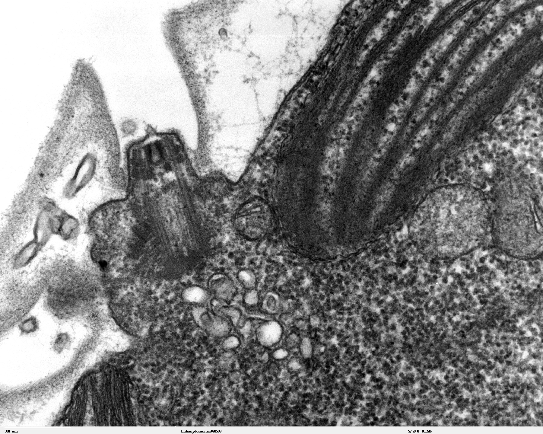
Transmission electron microscope image, showing an example of green algae (Chlorophyta). Chlamydomanas reinhardtii is a unicellular flagellate used as a model system in molecular genetics work and flagellar motility studies. This image shows the flagellar apparatus, just after flagellar excision, which occurs at the transition zone(see area of flagella, with its fibers of the stellate structure). This image also shows components of the contractile vacuoles which are located just below the flagellar apparatus. JEOL 100CX TEM |
| 日付 | |
| 原典 |
Source and public domain notice at: |
| 作者 | Elizabeth Smith, Louisa Howard, Erin Dymek (Dartmouth Electron Microscope Facility, Dartmouth College) |
| 許可 (ファイルの再利用) |
Released into the public domain |
ライセンス
| この著作物は、著作者であるElizabeth Smith, Louisa Howard and Erin Dymekによって権利が放棄され、パブリックドメインとされました。これは全世界で適用されます。 一部の国では、これが法的に可能ではない場合があります。その場合は、次のように宣言します。 Elizabeth Smith, Louisa Howard and Erin Dymekは、あらゆる人に対して、法により必要とされている条件を除き、如何なる条件も課すことなく、あらゆる目的のためにこの著作物を使用する権利を与えます。
|
ファイルの履歴
過去の版のファイルを表示するには、その版の日時をクリックしてください。
| 日付と時刻 | サムネイル | 寸法 | 利用者 | コメント | |
|---|---|---|---|---|---|
| 現在の版 | 2006年10月7日 (土) 14:03 | | 1,800 × 1,438 (1,010キロバイト) | Patho | {{Information |Description=Transmission electron microscope image, showing an example of green algae (Chlorophyta). Chlamydomanas reinhardtii is a unicellular flagellate used as a model system in molecular genetics work and flagellar motility studies. T |
ファイルの使用状況
以下のページがこのファイルを使用しています:
グローバルなファイル使用状況
以下に挙げる他のウィキがこの画像を使っています:
- de.wikibooks.org での使用状況
- es.wikipedia.org での使用状況
- vi.wikipedia.org での使用状況