ファイル:Mycobacterium tuberculosis.jpg
元のファイル (1,997 × 1,927 ピクセル、ファイルサイズ: 543キロバイト、MIME タイプ: image/jpeg)
ウィキメディア・コモンズのファイルページにある説明を、以下に表示します。
|
概要
| 解説 |
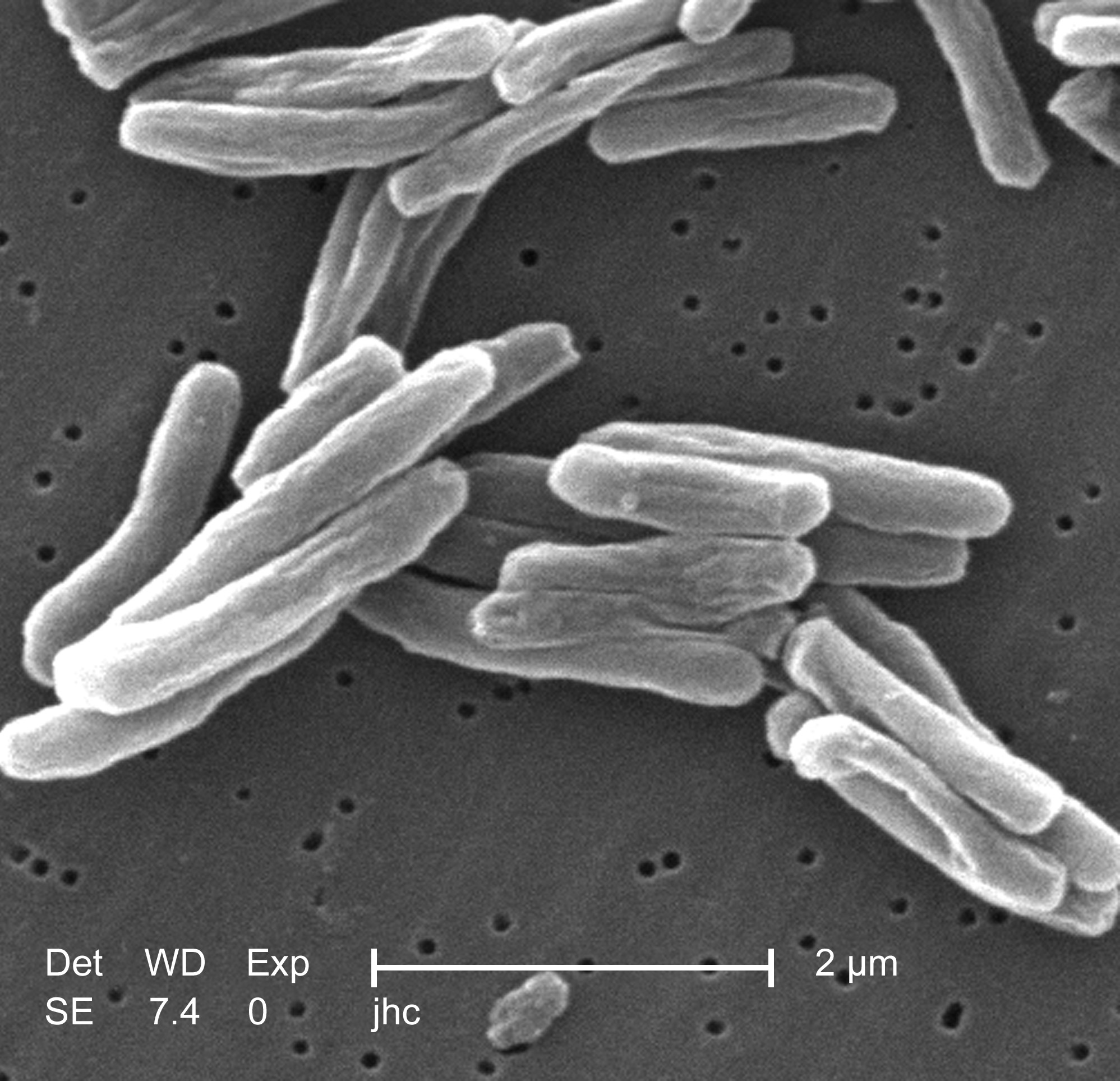
English: Under a high magnification of 15549x, this scanning electron micrograph (SEM) depicted some of the ultrastructural details seen in the cell wall configuration of a number of Gram-positive Mycobacterium tuberculosis bacteria. As an obligate aerobic organism M. tuberculosis can only survive in an environment containing oxygen. This bacterium ranges in length between 2 - 4 microns, and a width between 0.2 - 0.5 microns. See PHIL 9997 for a colorized version of this image.
TB bacteria become active, and begin to multiply, if the immune system can't stop them from growing. The bacteria attack the body and destroy tissue. If in the lungs, the bacteria can actually create a hole in the lung tissue. Some people develop active TB disease soon after becoming infected, before their immune system can fight off the bacteria. Other people may get sick later, when their immune system becomes weak for another reason. Babies and young children often have weak immune systems. People infected with HIV, the virus that causes AIDS, have very weak immune systems. Other people can have weak immune systems, too, especially people with any of these conditions: substance abuse; diabetes mellitus; silicosis; cancer of the head or neck; leukemia or Hodgkin's disease; severe kidney disease; low body weight; certain medical treatments (such as corticosteroid treatment or organ transplants); specialized treatment for rheumatoid arthritis, or Crohn's diseaseFrançais : Mycobacterium tuberculosis grossi 15 549 fois.
Español: Mycobacterium tuberculosis ampliado a 15549x.
中文:掃描電子顯微鏡下的結核桿菌.
Suomi: Mycobacterium tuberculosis 15549-kertaisena suurennoksena.
Čeština: Bakterie Mycobacterium tuberculosis, původce TBC.
Magyar: Mycobacterium tuberculosis.
한국어: 결핵균의 전자현미경 사진.
Simple English: TB Bacteria.
Kurdî: Girtineke elektronmîkroskobîk a bakteriyên tûberkûlozê pêk tînin.
Afrikaans: 'n Skanderende mikrograaf van Mycobacterium tuberculosis.
粵語: 掃描電子顯微鏡下嘅結核桿菌. |
||
| 日付 | |||
| 原典 |
|
||
| 作者 |
|
||
| 許可 (ファイルの再利用) |
PD-USGov-HHS-CDC English: None - This image is in the public domain and thus free of any copyright restrictions. As a matter of courtesy we request that the content provider be credited and notified in any public or private usage of this image. |
||
| その他のバージョン |
|

ライセンス
この画像は、アメリカ合衆国保健福祉省の一部である疾病予防管理センターの著作物であり、職員の公務の一環として撮影または作成されたものです。アメリカ合衆国連邦政府の著作物として、画像はパブリックドメインの状態にあります。
|
元のアップロードログ
- 2006-10-06 23:04 TimVickers 220×212×8 (10514 bytes) Janice Carr, CDC, http://www.cbc.ca/health/story/2006/03/17/tb-who060317.html?ref=rss
ファイルの履歴
過去の版のファイルを表示するには、その版の日時をクリックしてください。
| 日付と時刻 | サムネイル | 寸法 | 利用者 | コメント | |
|---|---|---|---|---|---|
| 現在の版 | 2019年6月21日 (金) 15:31 | | 1,997 × 1,927 (543キロバイト) | Tholme | Higher resolution |
| 2008年7月9日 (水) 00:53 |  | 220 × 212 (10キロバイト) | Reborned | {{Information |Description={{en|Janice Carr, CDC, http://www.cbc.ca/health/story/2006/03/17/tb-who060317.html?ref=rss}} |Source=Transferred from [http://en.wikipedia.org en.wikipedia] |Date=2006-10-06 (original upload date) |Author=Original uploader was [ |
ファイルの使用状況
以下のページがこのファイルを使用しています:
グローバルなファイル使用状況
以下に挙げる他のウィキがこの画像を使っています:
- af.wikipedia.org での使用状況
- ang.wikipedia.org での使用状況
- ar.wikipedia.org での使用状況
- arz.wikipedia.org での使用状況
- as.wikipedia.org での使用状況
- bcl.wikipedia.org での使用状況
- bg.wikipedia.org での使用状況
- bn.wikipedia.org での使用状況
- bo.wikipedia.org での使用状況
- bs.wikipedia.org での使用状況
- ca.wikipedia.org での使用状況
- ckb.wikipedia.org での使用状況
- cs.wikipedia.org での使用状況
- da.wikipedia.org での使用状況
- el.wikipedia.org での使用状況
- en.wikipedia.org での使用状況
- Disease
- Robert Koch
- Tuberculosis
- Mantoux test
- Pott's disease
- Health in China
- Wikipedia:Selected anniversaries/March 24
- Heaf test
- Tine test
- Tuberculosis radiology
- Tuberculosis diagnosis
- Tuberculin
- Chest photofluorography
- Tuberculosis verrucosa cutis
- The Global Fund to Fight AIDS, Tuberculosis and Malaria
- Latent tuberculosis
- Cultural depictions of tuberculosis
- Auramine phenol stain
- Interferon gamma release assay
- Mycobacterium caprae
このファイルのグローバル使用状況を表示する。