ファイル:Nematocyst discharge.png
高解像度版はありません。
Nematocyst_discharge.png (480 × 371 ピクセル、ファイルサイズ: 190キロバイト、MIME タイプ: image/png)
ウィキメディア・コモンズのファイルページにある説明を、以下に表示します。
|

|
この人体や生物に関する画像は、ベクターイメージである SVG ファイルとして再作成されるべきです。これにはいくつかの利点があります。詳しくはCommons:Media for cleanupを参照してください。この画像の SVG 形式がすでに利用可能である場合は、アップロードしてください。アップロード後、この画像にあるこのテンプレートを{{Vector version available|新しい画像ファイル名.svg}}テンプレートで置き換えてください。
|
概要
| 解説 |
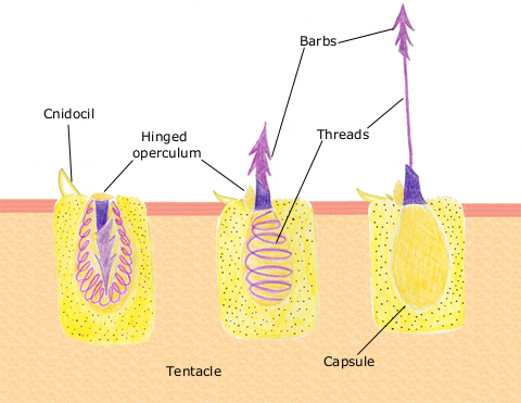
English: The diagram above shows the anatomy of a nematocyst cell and its “firing” sequence, from left to right. On the far left is a nematocyst inside its cellular capsule. The cell’s thread is coiled under pressure and wrapped around a stinging barb. When potential prey makes contact with the tentacles of a polyp, the nematocyst cell is stimulated. This causes a flap of tissue covering the nematocyst—the operculum—to fly open. The middle image shows the open operculum, the rapidly uncoiling thread and the emerging barb. On the far right is the fully extended cell. The barbs at the end of the nematocyst are designed to stick into the polyp’s victim and inject a poisonous liquid. When subdued, the polyp’s tentacles move the prey toward its mouth and the nematocysts recoil back into their capsules. |
| 日付 | 2007年4月11日 (当初のアップロード日) |
| 原典 | en.wikipedia からコモンズに移動されました。 |
| 作者 | オリジナルのアップロード者は英語版ウィキペディアのSpaullyさん |
ライセンス
This file is licensed under Creative Commons ShareAlike 1.0 License.
Creative Commons has retired this legal tool and does not recommend that it be applied to works.
|
|
|
元のアップロードログ
元のファイルページはこちら。以下の利用者は全てen.wikipediaに属します。
- 2007-04-11 17:10 Spaully 480×371×8 (194868 bytes) Modified from: http://www.oceanservice.noaa.gov/education/kits/corals/media/supp_coral01b.html {{Information |Description=Nematocyst discharge process. |Source= Modified from [http://www.oceanservice.noaa.gov/education/kits/corals/media/supp_coral01b.html
ファイルの履歴
過去の版のファイルを表示するには、その版の日時をクリックしてください。
| 日付と時刻 | サムネイル | 寸法 | 利用者 | コメント | |
|---|---|---|---|---|---|
| 現在の版 | 2007年10月13日 (土) 17:29 | | 480 × 371 (190キロバイト) | Alison | {{Information |Description===Description== The diagram above shows the anatomy of a nematocyst cell and its “firing” sequence, from left to right. On the far left is a nematocyst inside its cellular capsule. The cell’s thread is coiled under pressur |
ファイルの使用状況
以下のページがこのファイルを使用しています:
グローバルなファイル使用状況
以下に挙げる他のウィキがこの画像を使っています:
- ca.wikipedia.org での使用状況
- ceb.wikipedia.org での使用状況
- en.wikipedia.org での使用状況
- fr.wikipedia.org での使用状況
- hr.wikipedia.org での使用状況
- id.wikipedia.org での使用状況
- it.wikibooks.org での使用状況
- lv.wikipedia.org での使用状況
- ms.wikipedia.org での使用状況
- my.wikipedia.org での使用状況
- pa.wikipedia.org での使用状況
- pt.wikipedia.org での使用状況
- simple.wikipedia.org での使用状況
- sv.wikipedia.org での使用状況
- te.wikipedia.org での使用状況
- th.wikipedia.org での使用状況
- vi.wikipedia.org での使用状況