ファイル:Siphovirus.tiff

この TIF ファイルのこの JPG プレビューのサイズ: 594 × 599 ピクセル. その他の解像度: 238 × 240 ピクセル | 476 × 480 ピクセル | 761 × 768 ピクセル | 1,048 × 1,057 ピクセル。
元のファイル (1,048 × 1,057 ピクセル、ファイルサイズ: 1.09メガバイト、MIME タイプ: image/tiff)
ウィキメディア・コモンズのファイルページにある説明を、以下に表示します。
|
概要
| 解説 |
English: Electron micrographs of bacteriophages from Cutibacterium acnes (formerly Propionibacterium acnes). Phages were negatively stained with 0.75% uranyl formate and subjected to transmission electron microscopy. The phages have a head of approximately 55 nm in diameter, loaded with genetic material. Their tails have a size of 150 × 10 nm and are flexible and non-contractile. The upper micrographs show Propionibacterium phage PAD40 and PAD11 (NBBI TaxIDs 504513 and 504513 respectively). |
| 日付 | |
| 原典 | BMC Microbiology 2008, 8:139 doi:10.1186/1471-2180-8-139 |
| 作者 | Rolf Lood, Matthias Mörgelin, Anna Holmberg, Magnus Rasmussen and Mattias Collin |
ライセンス
このファイルはクリエイティブ・コモンズ 表示 2.5 一般ライセンスのもとに利用を許諾されています。
- あなたは以下の条件に従う場合に限り、自由に
- 共有 – 本作品を複製、頒布、展示、実演できます。
- 再構成 – 二次的著作物を作成できます。
- あなたの従うべき条件は以下の通りです。
- 表示 – あなたは適切なクレジットを表示し、ライセンスへのリンクを提供し、変更があったらその旨を示さなければなりません。これらは合理的であればどのような方法で行っても構いませんが、許諾者があなたやあなたの利用行為を支持していると示唆するような方法は除きます。
ファイルの履歴
過去の版のファイルを表示するには、その版の日時をクリックしてください。
| 日付と時刻 | サムネイル | 寸法 | 利用者 | コメント | |
|---|---|---|---|---|---|
| 現在の版 | 2011年12月2日 (金) 14:31 | 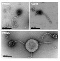 | 1,048 × 1,057 (1.09メガバイト) | Alexbateman |
ファイルの使用状況
以下のページがこのファイルを使用しています:
グローバルなファイル使用状況
以下に挙げる他のウィキがこの画像を使っています:
- ar.wikipedia.org での使用状況
- arz.wikipedia.org での使用状況
- ca.wikipedia.org での使用状況
- de.wikipedia.org での使用状況
- en.wikipedia.org での使用状況
- it.wikipedia.org での使用状況
- www.mediawiki.org での使用状況
- www.wikidata.org での使用状況
- zh.wikipedia.org での使用状況