ファイル:Chlamydomanas reinhardtii Flagella 5 - TEM.jpg

元のファイル (1,800 × 1,438 ピクセル、ファイルサイズ: 650キロバイト、MIME タイプ: image/jpeg)
ウィキメディア・コモンズのファイルページにある説明を、以下に表示します。
|
概要
| 解説 |
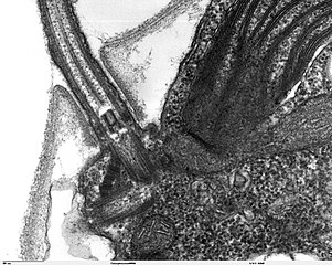
Transmission electron microscope image, showing an example of green algae (Chlorophyta). Chlamydomanas reinhardtii is a unicellular flagellate used as a model system in molecular genetics work and flagellar motility studies. This image is a longitudinal section through a portion of the flagellar apparatus. In the cell apex are the basal body regions that are the anchoring sites for the flagella. This image shows that the two flagella form a V and they are connected at their bases by a transversely striated fibre. This connection is thought to play a part in the coordination of flagellar movement. Also visible is the transition region, with its fibers of the stellate structure. JEOL 100CX TEM |
| 原典 | |
| 作者 | Elizabeth Smith, Louisa Howard, Erin Dymek |
| 許可 (ファイルの再利用) |
PD |
ライセンス
| この著作物は、著作者であるElizabeth Smith, Louisa Howard and Erin Dymekによって権利が放棄され、パブリックドメインとされました。これは全世界で適用されます。 一部の国では、これが法的に可能ではない場合があります。その場合は、次のように宣言します。 Elizabeth Smith, Louisa Howard and Erin Dymekは、あらゆる人に対して、法により必要とされている条件を除き、如何なる条件も課すことなく、あらゆる目的のためにこの著作物を使用する権利を与えます。
|
ファイルの履歴
過去の版のファイルを表示するには、その版の日時をクリックしてください。
| 日付と時刻 | サムネイル | 寸法 | 利用者 | コメント | |
|---|---|---|---|---|---|
| 現在の版 | 2006年10月7日 (土) 14:01 | | 1,800 × 1,438 (650キロバイト) | Patho | {{Information |Description=Transmission electron microscope image, showing an example of green algae (Chlorophyta). Chlamydomanas reinhardtii is a unicellular flagellate used as a model system in molecular genetics work and flagellar motility studies. T |
ファイルの使用状況
グローバルなファイル使用状況
以下に挙げる他のウィキがこの画像を使っています:
- de.wikibooks.org での使用状況
- uk.wikipedia.org での使用状況