ファイル:Chlamydomonas TEM 09.jpg

このプレビューのサイズ: 751 × 600 ピクセル。 その他の解像度: 301 × 240 ピクセル | 601 × 480 ピクセル | 961 × 768 ピクセル | 1,280 × 1,023 ピクセル | 1,800 × 1,438 ピクセル。
元のファイル (1,800 × 1,438 ピクセル、ファイルサイズ: 784キロバイト、MIME タイプ: image/jpeg)
ウィキメディア・コモンズのファイルページにある説明を、以下に表示します。
|
| 解説 |
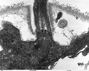
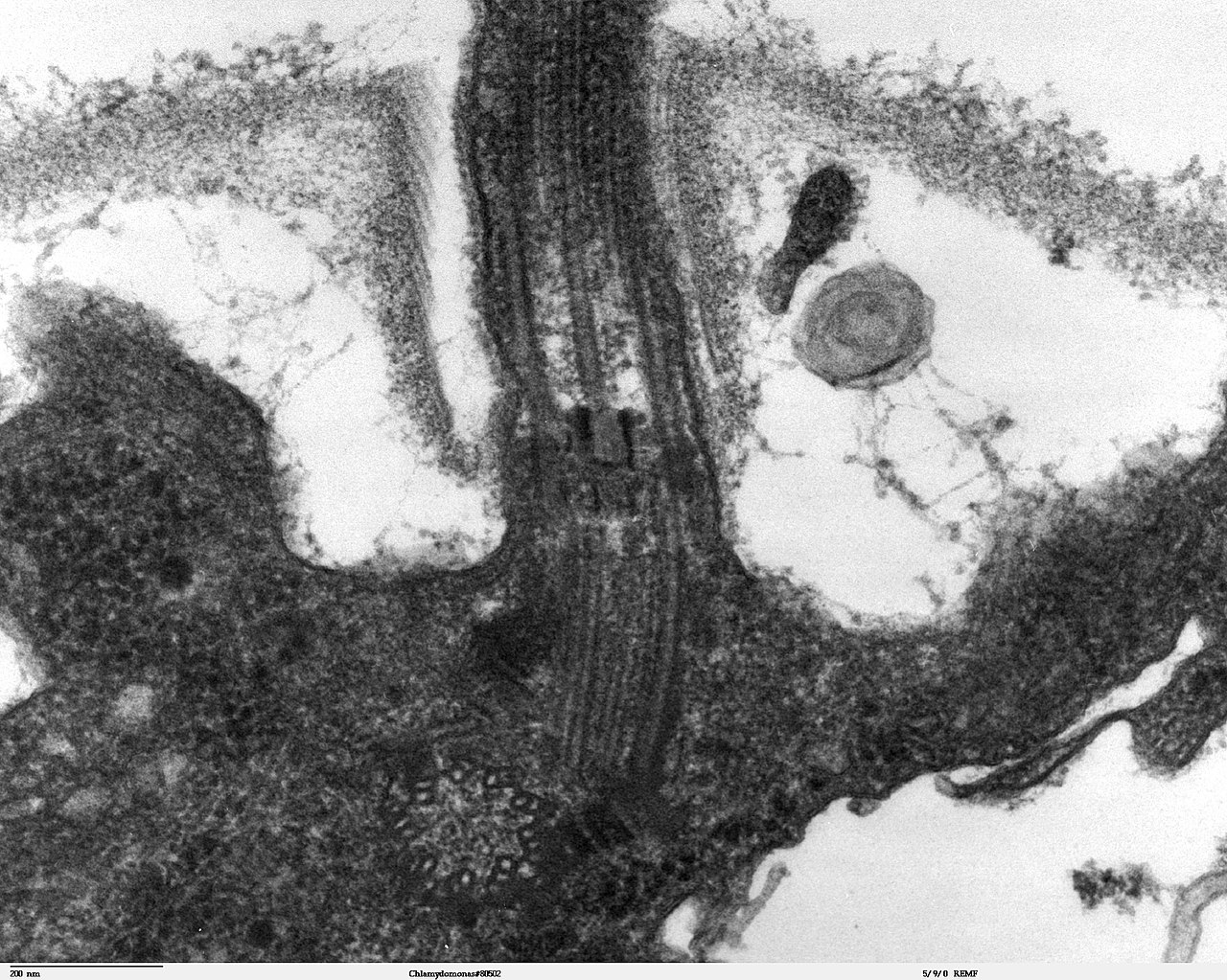
Transmission electron microscope image, showing an example of green algae (Chlorophyta). Chlamydomanas reinhardtii is a unicellular flagellate used as a model system in molecular genetics work and flagellar motility studies. This image is a longitudinal section through the flagella area. In the cell apex is the basal body that is the anchoring site for a flagella. Basal bodies originate from and have a substructure similar to that of centrioles, with nine peripheral microtubule triplets(see structure at bottom center of image). The two inner microtubules of each triplet in a basal body become the two outer doublets in the flagella. This image also shows the transition region, with its fibers of the stellate structure. The top of the image shows the flagella passing through the cell wall. |
| 日付 | |
| 原典 | Source and public domain notice at http://remf.dartmouth.edu/imagesindex.html |
| 作者 | Dartmouth Electron Microscope Facility, Dartmouth College |
| 許可 (ファイルの再利用) |
Released into the public domain |
| この著作物は、著作者であるDartmouth Electron Microscope Facility, Dartmouth Collegeによって権利が放棄され、パブリックドメインとされました。これは全世界で適用されます。 一部の国では、これが法的に可能ではない場合があります。その場合は、次のように宣言します。 Dartmouth Electron Microscope Facility, Dartmouth Collegeは、あらゆる人に対して、法により必要とされている条件を除き、如何なる条件も課すことなく、あらゆる目的のためにこの著作物を使用する権利を与えます。
|
ファイルの履歴
過去の版のファイルを表示するには、その版の日時をクリックしてください。
| 日付と時刻 | サムネイル | 寸法 | 利用者 | コメント | |
|---|---|---|---|---|---|
| 現在の版 | 2007年9月21日 (金) 06:47 | | 1,800 × 1,438 (784キロバイト) | Neil916 | {{Information |Description= Transmission electron microscope image, showing an example of green algae (Chlorophyta). <br><br>''Chlamydomanas reinhardtii'' is a unicellular flagellate used as a model system in molecular genetics work and flagellar motilit |
ファイルの使用状況
グローバルなファイル使用状況
以下に挙げる他のウィキがこの画像を使っています:
- ar.wikipedia.org での使用状況
- bs.wikipedia.org での使用状況
- ca.wikipedia.org での使用状況
- cs.wikipedia.org での使用状況
- de.wikipedia.org での使用状況
- de.wikibooks.org での使用状況
- en.wikipedia.org での使用状況
- es.wikipedia.org での使用状況
- gl.wikipedia.org での使用状況
- id.wikipedia.org での使用状況
- ko.wikipedia.org での使用状況
- pl.wikipedia.org での使用状況
- ru.wikipedia.org での使用状況
- sv.wikipedia.org での使用状況
- tr.wikipedia.org での使用状況
- uk.wikipedia.org での使用状況
- zh.wikipedia.org での使用状況