鼻涙管
眼の涙嚢から鼻腔に涙を運ぶ管
鼻涙管(びるいかん、nasolacrimal duct, tear duct)は、眼の涙嚢から鼻腔に涙を運ぶ管[1][2]。この管は上顎骨と涙骨の間の眼窩から始まり、そこから下方と後方に向かって伸びる。鼻腔の下鼻道への鼻涙管の開口部は、粘膜ひだ(valve of Hasner or plica lacrimalis)で部分的に覆われている[3]。
| 鼻涙管 | |
|---|---|
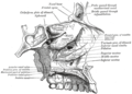
 右側の涙器 | |
 顔の骨の輪郭。含気腔の位置を示す。 | |
| 概要 | |
| 表記・識別 | |
| ラテン語 | Ductus nasolacrimalis |
| MeSH | D009301 |
| グレイ解剖学 | p.1029 |
| TA | A15.2.07.070 |
| FMA | 9703 |
| 解剖学用語 | |
余分な涙は鼻涙管を通って下鼻道へと流れる。泣いているときやアレルギーで涙目になっているときに鼻水が出たり目薬の味がしたりするのはこのためである。目薬を差すときに鼻涙管を指で押して閉じると薬が目から出て鼻腔に入り体のどこかに副作用が出るのを防ぐことができるのもこのためである。
臨床的意義 編集
鼻涙管閉塞が起こることがある[4][5][6]。これにより涙が過剰に出るEpiphora (medicine)(慢性低位鼻涙管閉塞症)が起こる[7]。先天的な閉塞があると、管が嚢胞状に拡大することがあり、これは涙嚢ヘルニアまたは Timo cyst と呼ばれる。ドライアイの人は涙管を塞いで液体の排出量を制限し、水分を保持するために涙点プラグを装着することができる。
耳の感染症では、過剰な粘液が涙とは逆に鼻涙管を通って排出されることがある[要出典]。
ヒトでは、男性の涙管は女性の涙管よりも大きい傾向がある[8]。
他の画像 編集
-
 左鼻腔
左鼻腔

関連項目 編集
出典 編集
- ^ Nasolacrimal System Anatomy: Embryology, Puncta, Canaliculi. (2020-02-19).
- ^ Herbert, Ronald A.; Janardhan, Kyathanahalli S.; Pandiri, Arun R.; Cesta, Mark F.; Miller, Rodney A. (2018). “Nose, Larynx, and Trachea”. Boorman's Pathology of the Rat. Elsevier. pp. 391–435. doi:10.1016/b978-0-12-391448-4.00022-8. ISBN 978-0-12-391448-4. "The paired nasolacrimal ducts carry lacrimal secretions from the eye to the nasal cavity, and originate as oval openings near the edge of the medial canthus of the eyelids. Initially the duct is small and circular, but in the middle portion, the diameter increases and the appearance is more oblong and saccular. The diameter again decreases before the duct enters the ventrolateral nasal vestibule medial to the root of the incisor tooth approximately 2 mm caudal to the nares."
- ^ “Medical Definition of PLICA LACRIMALIS”. Merriam-Webster Medical Dictionary (2020年5月8日). 2020年5月8日閲覧。 “an imperfect valve at the opening of the nasolacrimal duct into the inferior meatus of the nose”
- ^ CARREIRO, J (2009). “Head and neck”. Pediatric Manual Medicine. Elsevier. pp. 13–97. doi:10.1016/b978-0-443-10308-7.00002-8. ISBN 978-0-443-10308-7. "Dacryostenosis is a condition whereby the nasolacrimal duct is narrowed or blocked and the glandular secretions into the eye are prevented from draining properly. The secretions collect around the orifice of the duct and in the corner of the eye, where they thicken, resulting in a gooey, sticky substance that further clogs any drainage route."
- ^ Stagner, Anna M.; Jakobiec, Frederick A.; Eagle, Ralph C.; Charles, Norman C. (2018). “Infections of the Eye and Its Adnexa”. Diagnostic Pathology of Infectious Disease. Elsevier. pp. 648–685. doi:10.1016/b978-0-323-44585-6.00021-7. ISBN 978-0-323-44585-6. "Dacryocystitis is produced by obstruction of the nasolacrimal duct with resultant tear stasis. Clinically, patients display erythema and swelling of the lacrimal sac, creating a mass in the medial canthal area, centered below the medial canthal tendon."
- ^ "Blocked tear ducts in infants" Archived 2012-07-08 at the Wayback Machine., Pediatric Views, June 2006.
- ^ Nerad, Jeffrey A.; Carter, Keith D.; Alford, Mark (2008). “Disorders of the Lacrimal System: Congenital Obstruction”. Oculoplastic and Reconstructive Surgery. Elsevier. pp. 131–137. doi:10.1016/b978-0-323-05386-0.50010-7. ISBN 978-0-323-05386-0
- ^ "Tears of Men and Women Are Different", Wall Street Journal, mirrored at "Why Men and Women Shed Different Tears", Fox News, May 5, 2011.
外部リンク 編集
- Anatomy figure: 33:04-09 at Human Anatomy Online, SUNY Downstate Medical Center
- lesson9 at The Anatomy Lesson by Wesley Norman (Georgetown University)