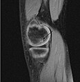
ファイル:OCD WalterReed MRI-Sagital-T1.jpeg

このプレビューのサイズ: 579 × 600 ピクセル。 その他の解像度: 232 × 240 ピクセル | 463 × 480 ピクセル | 945 × 979 ピクセル。
元のファイル (945 × 979 ピクセル、ファイルサイズ: 353キロバイト、MIME タイプ: image/jpeg)
ファイルの履歴
過去の版のファイルを表示するには、その版の日時をクリックしてください。
| 日付と時刻 | サムネイル | 寸法 | 利用者 | コメント | |
|---|---|---|---|---|---|
| 現在の版 | 2009年3月4日 (水) 21:10 |  | 945 × 979 (353キロバイト) | FoodPuma | Added arrow (edited with Adobe Photoshop CS2) |
| 2009年2月8日 (日) 00:12 |  | 516 × 467 (79キロバイト) | FoodPuma | {{Information |Description={{en|1="Sagittal and coronal T1 and T2 images demonstrate linear low T1, high T2 signal at the articular surfaces of the lateral aspects of the medial femoral condyles bilaterally, corresponding to the radiographs, confirming th |
ファイルの使用状況
以下のページがこのファイルを使用しています:
グローバルなファイル使用状況
以下に挙げる他のウィキがこの画像を使っています:
- ar.wikipedia.org での使用状況
- az.wikipedia.org での使用状況
- ca.wikipedia.org での使用状況
- en.wikipedia.org での使用状況
- nl.wikipedia.org での使用状況
- pl.wikipedia.org での使用状況
- uk.wikipedia.org での使用状況
- www.wikidata.org での使用状況
- zh.wikipedia.org での使用状況
